reedley-cerec-machine-1200x488.jpg

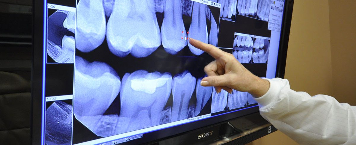
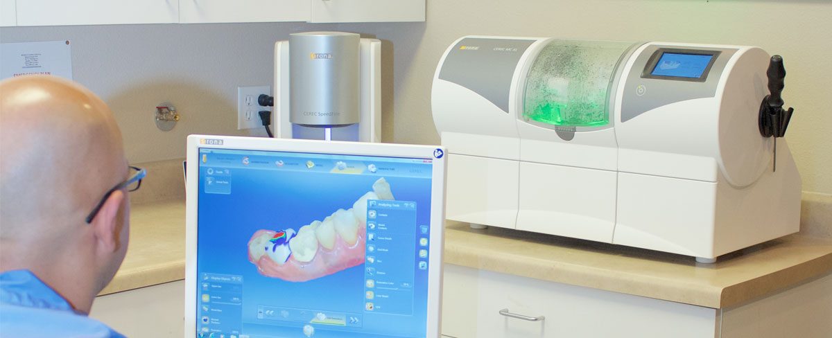
CEREC stands for “Chairside Economical Restoration of Esthetic Ceramics” and is a computer-aided design and manufacturing system for dentists. CEREC combines a camera, computer, and milling instrument into one machine allowing tooth restorations to be created in the dentist office, all within a single visit.

Not only does this eliminate both the long process of sending your tooth mold to an outside lab and living with a temporary crown, hundreds of studies show that the CEREC system is a safer and more effective way to restore your teeth than traditional methods.

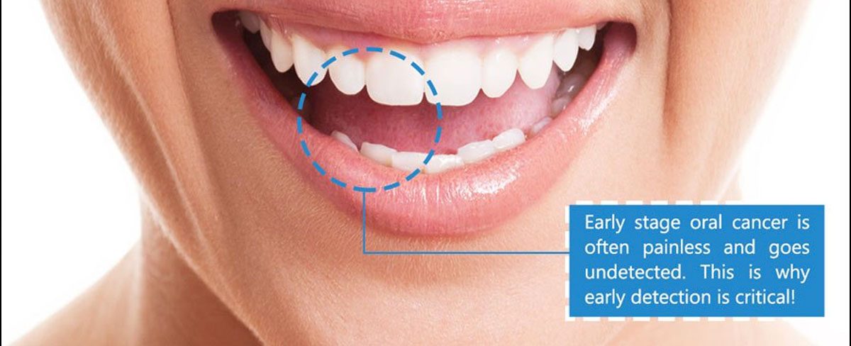
The CEREC system is used to fix damaged or unsightly teeth. Whether you’re looking for a more radiant smile, or you’re teeth are decaying and need to be repaired, CEREC can help.

How CEREC works

CEREC uses advanced computer-aided design technology and software to create perfect, natural-looking restorations in just minutes. Thanks to this technology, visiting your dentist for a tooth restoration is a simple and non-invasive procedure.

Here’s how it works:

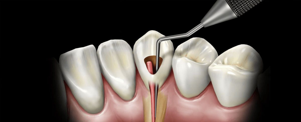
  • Your dentist will prepare your tooth for the restoration
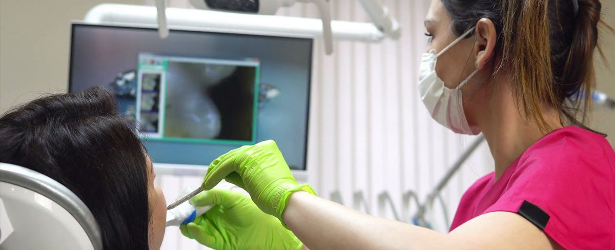
  • The CEREC software takes a digital photo of the treatment area
  • The CEREC technology converts the image into a 3D virtual model
  • With input from the dentist, the software creates the final restoration
  • The CEREC mill precisely carves a ceramic block into the exact shape and specifications of your restoration
  • Once the dentist has ensured the restoration fits properly in your mouth, the restoration is polished and bonded to your tooth

This entire process takes no longer than 2 hours!

If you have any questions or would like to schedule an introductory visit, please give our office a call at: (559) 637-0123.


oral-cancer-1200x488.jpg

Oral cancer screening is an examination performed by a dentist or doctor to look for signs of cancer or precancerous conditions in your mouth.

The goal of oral cancer screening is to identify mouth cancer early, when there is a greater chance for a cure.

Most dentists perform an examination of your mouth during a routine dental visit to screen for oral cancer. Some dentists may use additional tests to aid in identifying areas of abnormal cells in your mouth.

Medical organizations disagree on whether healthy people without risk factors for mouth cancer need oral cancer screening. No single oral exam or oral cancer screening test is proved to reduce the risk of dying of oral cancer. Still, you and your dentist may decide that an oral exam or a special test is right for you based on your risk factors.

Why it’s done

The goal of oral cancer screening is to detect mouth cancer or precancerous lesions that may lead to mouth cancer at an early stage — when cancer or lesions are easiest to remove and most likely to be cured.

But no studies have proved that oral cancer screening saves lives, so not all organizations agree about the benefits of an oral exam for oral cancer screening. Some groups recommend screening, while others don’t.

People with a high risk of oral cancer may be more likely to benefit from oral cancer screening, though studies haven’t clearly proved that. Factors that can increase the risk of oral cancer include:

  • Tobacco use of any kind, including cigarettes, cigars, pipes, chewing tobacco and snuff, among others
  • Heavy alcohol use
  • Previous oral cancer diagnosis
  • History of significant sun exposure, which increases the risk of lip cancer

Ask Reedley Family Dental whether oral cancer screening is appropriate for you. Also ask about ways you can reduce your risk of oral cancer, such as quitting smoking and not drinking alcohol.


Rotary-Endodontics-1200x488.jpg

Root canal treatment, also called endodontic therapy, is necessary when the pulp of the tooth is infected following significant caries, cracks in the enamel or a trauma (shock). If the inflamed or infected pulp is left untreated, it can cause pain or lead to an abscess. This treatment involves the removal of the infected pulp, the disinfection, and the filling and sealing of the root canal system. The tooth is then restored with a permanent filling or a crown.

To clean the inside of the tooth, we use the combination of a mechanical method (instruments that remove the debris inside the pulp chamber and root canals) and a chemical method (desinfecting solution injected into the tooth to kill bacteria).

To remove the pulp from the root canals (mechanical method), the dentist uses precision instruments (endodontic files). These can be traditional manual files or electrically-powered instruments (rotary endodontic instruments).

Advantages of rotary endodontics

Rotary endodontic methods offer several advantages over manual root canal treatments:

  • Faster treatment (often in one appointment);
  • Patients feel much more comfortable during the treatment;
  • More reliable;
  • No unpleasant noises;
  • More precision (the flexibility of instruments allows a better negotiation of curved canals);
  • Higher quality treatments (more consistent).

Intraoral-Camera-1200x488.jpg

The short answer is that an intraoral camera is a tool our dentist at Reedley Family Dental uses to examine your mouth in as detailed a way as possible. The instrument, which may look like an oversized pen, has a camera that takes high-resolution footage or images of a patient’s mouth and shows the visuals real-time on a monitor—they’re like high-tech versions of the hand mirrors you see in your dentist’s practice.

Benefits the dentist.

Intraoral cameras have incredible technological features. With LED lighting, a head that rotates from 0 to 90 degrees, and powerful magnifying capabilities (some cameras can zoom in up to 100x), our dentist can examine your mouth in extreme detail. This means he or she can make diagnoses more accurately. The office can attach these photos to your health record to make tracking any changes simple. Additionally, because the visuals from the intraoral camera appear on the monitor as they’re taken, our dentist can discuss your oral health with you while you both see the images or footage.

Benefits the patient.

Each feature that benefits the dentist also benefits the patient—maybe even more. Our dentist understands symptoms and conditions thoroughly, but it’s often difficult to explain precisely what is happening in a patient’s mouth using just a mouth mirror, which is small and hard to see, or an x-ray image, which takes time to print and doesn’t display images clearly.

When our dentist uses an intraoral camera during your examination, however, you’re seeing exactly what he or she sees right then. Dentists can display clear, colorful images, allowing them to point out any issues and discuss them with you immediately. You’ll certainly learn a lot about your mouth! And the more you see and understand, the more confident you can be when making treatment decisions.

At Reedley Family Dental, we will print or email images for our patients so you can refer to them at home. Because these images are processed immediately, it saves you time.


digital-dental-x-ray-1200x488.jpg

Our natural environment exposes us to all kinds of radiation. Digital x-rays produce a significantly lower level of radiation compared to older methods. Not only are digital x-rays faster and better for your health, they are also more comfortable. For our new patients, we generally recommend a full mouth series and bitewing x-rays. These are a good reference for 3-5 years and are usually taken after your check-up and cleaning.

5 reasons for taking X-Rays:

  1. To look for decay in between teeth
  2. To check for the bone loss associated with gum disease
  3. To check for decay under older fillings
  4. To see if there are signs of infection at the very bottom of the tooth root
  5. To examine a problem area before it is treated with a procedure

Panorex-1200x488.jpg

Panoramic dental x-ray uses a very small dose of ionizing radiation to capture the entire mouth in one image. It is commonly performed by dentists and oral surgeons in everyday practice and may be used to plan treatment for dentures, braces, extractions, and implants.

This exam requires little to no special preparation. Tell your doctor if there is a possibility you are pregnant. Remove any jewelry, eyeglasses or metal objects that might interfere with the x-ray images. You will be asked to wear a lead apron to protect the rest of your body from any radiation exposure.

What are some common uses of the procedure?

A panoramic x-ray is a commonly performed examination by dentists and oral surgeons in everyday practice and is an important diagnostic tool. It covers a wider area than a conventional intraoral x-ray and, as a result, provides valuable information about the maxillary sinuses, tooth positioning, and other bone abnormalities. This examination is also used to plan treatment for full and partial dentures, braces, extractions, and implants.

A panoramic x-ray can also reveal dental and medical problems such as:

  • advanced periodontal disease
  • cysts in the jaw bones
  • jaw tumors and oral cancer
  • impacted teeth including wisdom teeth
  • jaw disorders (also known as temporomandibular joint or TMJ disorders)
  • sinusitis

What are the benefits vs. risks?

Benefits

  • No radiation remains in a patient’s body after an x-ray examination.
  • X-rays usually have no side effects in the typical diagnostic range for this exam.
  • Panoramic x-rays can be used for very young children since the film does not have to be placed inside the mouth.

Risks

  • Women should always inform their dentist or oral surgeon if there is any possibility that they are pregnant.

Planmeca-brand-Promax-3D-1200x488.jpg

Dental cone beam computed tomography (CT) is a special type of x-ray equipment used when regular dental or facial x-rays are not sufficient. Our Dentist may use this technology to produce three dimensional (3-D) images of your teeth, soft tissues, nerve pathways and bone in a single scan.

This procedure requires little to no special preparation. Tell your doctor if there’s a possibility you are pregnant. Wear loose, comfortable clothing and leave jewelry at home. You may be asked to wear a gown.

What are some common uses of the procedure?

Dental cone beam CT is commonly used for treatment planning of orthodontic issues. It is also useful for more complex cases that involve:

  • surgical planning for impacted teeth.
  • diagnosing temporomandibular joint disorder (TMJ).
  • accurate placement of dental implants.
  • evaluation of the jaw, sinuses, nerve canals and nasal cavity.
  • detecting, measuring and treating jaw tumors.
  • determining bone structure and tooth orientation.
  • locating the origin of pain or pathology.
  • cephalometric analysis.
  • reconstructive surgery.

Copyright 2020 - Reedley Family Dental. Web Design & SEO by Tommy Le Inc.