Rotary-Endodontics-1200x488.jpg

Root canal treatment, also called endodontic therapy, is necessary when the pulp of the tooth is infected following significant caries, cracks in the enamel or a trauma (shock). If the inflamed or infected pulp is left untreated, it can cause pain or lead to an abscess. This treatment involves the removal of the infected pulp, the disinfection, and the filling and sealing of the root canal system. The tooth is then restored with a permanent filling or a crown.

To clean the inside of the tooth, we use the combination of a mechanical method (instruments that remove the debris inside the pulp chamber and root canals) and a chemical method (desinfecting solution injected into the tooth to kill bacteria).

To remove the pulp from the root canals (mechanical method), the dentist uses precision instruments (endodontic files). These can be traditional manual files or electrically-powered instruments (rotary endodontic instruments).

Advantages of rotary endodontics

Rotary endodontic methods offer several advantages over manual root canal treatments:

  • Faster treatment (often in one appointment);
  • Patients feel much more comfortable during the treatment;
  • More reliable;
  • No unpleasant noises;
  • More precision (the flexibility of instruments allows a better negotiation of curved canals);
  • Higher quality treatments (more consistent).

Intraoral-Camera-1200x488.jpg

The short answer is that an intraoral camera is a tool our dentist at Reedley Family Dental uses to examine your mouth in as detailed a way as possible. The instrument, which may look like an oversized pen, has a camera that takes high-resolution footage or images of a patient’s mouth and shows the visuals real-time on a monitor—they’re like high-tech versions of the hand mirrors you see in your dentist’s practice.

Benefits the dentist.

Intraoral cameras have incredible technological features. With LED lighting, a head that rotates from 0 to 90 degrees, and powerful magnifying capabilities (some cameras can zoom in up to 100x), our dentist can examine your mouth in extreme detail. This means he or she can make diagnoses more accurately. The office can attach these photos to your health record to make tracking any changes simple. Additionally, because the visuals from the intraoral camera appear on the monitor as they’re taken, our dentist can discuss your oral health with you while you both see the images or footage.

Benefits the patient.

Each feature that benefits the dentist also benefits the patient—maybe even more. Our dentist understands symptoms and conditions thoroughly, but it’s often difficult to explain precisely what is happening in a patient’s mouth using just a mouth mirror, which is small and hard to see, or an x-ray image, which takes time to print and doesn’t display images clearly.

When our dentist uses an intraoral camera during your examination, however, you’re seeing exactly what he or she sees right then. Dentists can display clear, colorful images, allowing them to point out any issues and discuss them with you immediately. You’ll certainly learn a lot about your mouth! And the more you see and understand, the more confident you can be when making treatment decisions.

At Reedley Family Dental, we will print or email images for our patients so you can refer to them at home. Because these images are processed immediately, it saves you time.


digital-dental-x-ray-1200x488.jpg

Our natural environment exposes us to all kinds of radiation. Digital x-rays produce a significantly lower level of radiation compared to older methods. Not only are digital x-rays faster and better for your health, they are also more comfortable. For our new patients, we generally recommend a full mouth series and bitewing x-rays. These are a good reference for 3-5 years and are usually taken after your check-up and cleaning.

5 reasons for taking X-Rays:

  1. To look for decay in between teeth
  2. To check for the bone loss associated with gum disease
  3. To check for decay under older fillings
  4. To see if there are signs of infection at the very bottom of the tooth root
  5. To examine a problem area before it is treated with a procedure

Panorex-1200x488.jpg

Panoramic dental x-ray uses a very small dose of ionizing radiation to capture the entire mouth in one image. It is commonly performed by dentists and oral surgeons in everyday practice and may be used to plan treatment for dentures, braces, extractions, and implants.

This exam requires little to no special preparation. Tell your doctor if there is a possibility you are pregnant. Remove any jewelry, eyeglasses or metal objects that might interfere with the x-ray images. You will be asked to wear a lead apron to protect the rest of your body from any radiation exposure.

What are some common uses of the procedure?

A panoramic x-ray is a commonly performed examination by dentists and oral surgeons in everyday practice and is an important diagnostic tool. It covers a wider area than a conventional intraoral x-ray and, as a result, provides valuable information about the maxillary sinuses, tooth positioning, and other bone abnormalities. This examination is also used to plan treatment for full and partial dentures, braces, extractions, and implants.

A panoramic x-ray can also reveal dental and medical problems such as:

  • advanced periodontal disease
  • cysts in the jaw bones
  • jaw tumors and oral cancer
  • impacted teeth including wisdom teeth
  • jaw disorders (also known as temporomandibular joint or TMJ disorders)
  • sinusitis

What are the benefits vs. risks?

Benefits

  • No radiation remains in a patient’s body after an x-ray examination.
  • X-rays usually have no side effects in the typical diagnostic range for this exam.
  • Panoramic x-rays can be used for very young children since the film does not have to be placed inside the mouth.

Risks

  • Women should always inform their dentist or oral surgeon if there is any possibility that they are pregnant.

Planmeca-brand-Promax-3D-1200x488.jpg

Dental cone beam computed tomography (CT) is a special type of x-ray equipment used when regular dental or facial x-rays are not sufficient. Our Dentist may use this technology to produce three dimensional (3-D) images of your teeth, soft tissues, nerve pathways and bone in a single scan.

This procedure requires little to no special preparation. Tell your doctor if there’s a possibility you are pregnant. Wear loose, comfortable clothing and leave jewelry at home. You may be asked to wear a gown.

What are some common uses of the procedure?

Dental cone beam CT is commonly used for treatment planning of orthodontic issues. It is also useful for more complex cases that involve:

  • surgical planning for impacted teeth.
  • diagnosing temporomandibular joint disorder (TMJ).
  • accurate placement of dental implants.
  • evaluation of the jaw, sinuses, nerve canals and nasal cavity.
  • detecting, measuring and treating jaw tumors.
  • determining bone structure and tooth orientation.
  • locating the origin of pain or pathology.
  • cephalometric analysis.
  • reconstructive surgery.

Alveoplasty-1200x488.jpg

Alveoplasty is the process of surgically re-contouring and modifying the jawbone ridge, usually after tooth extraction, to enhance the healing process and prepare the jawbone for subsequent dental restoration procedures such as the placement of dentures.

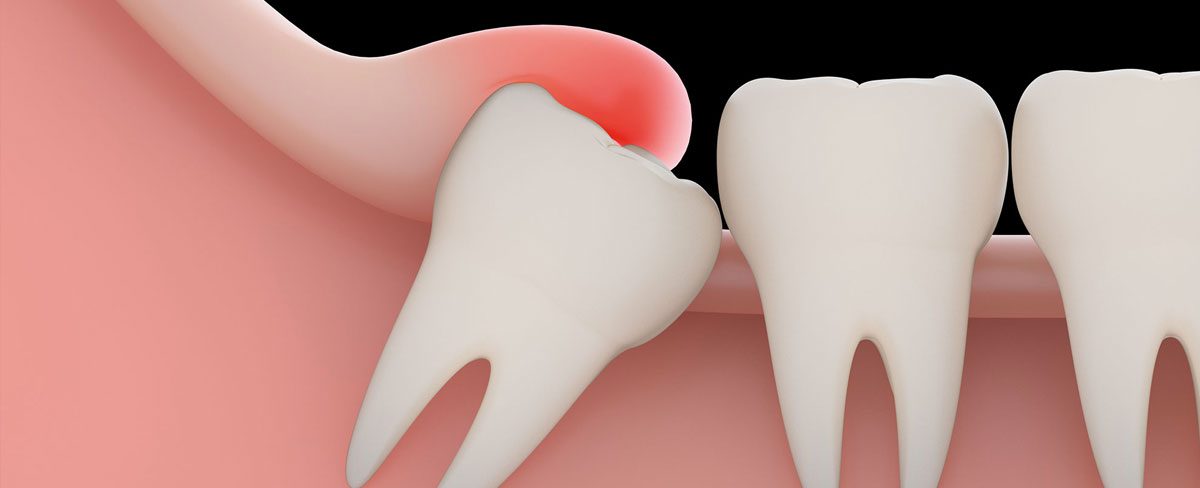
In some cases, tooth extraction leaves the jawbone surface uneven with high and low points in the socket where the tooth used to be. This poses a problem as the denture is likely to rub against the high points of the socket, making it unstable. Alveoplasty is performed in such cases to smooth out the jawbone to achieve correct alignment, which will make it easier for the dentist to fit dentures as well as help patients by enhancing retention of the said dental prosthesis.

Who Should Undergo and Expected Results

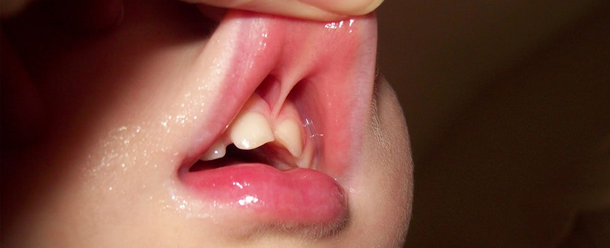
Alveoplasty can be recommended for patients following their tooth extraction to smooth out any rough bone ridges and remove any irregularities in the jawbone. This prepares the jawbone for the placement of dental prostheses such as dentures. The procedure can also be performed alone, without any preceding tooth extraction, for people who no longer have their complete set of natural teeth for a long time and have experienced weakness in their jawbone following bone loss. Some may even exhibit a ridge or lip of bone that protrudes.

This surgical procedure entails several days of rest, especially if performed with tooth extraction. The patient is placed on a soft diet for days to ensure the surgical wound remains undamaged and unopened.

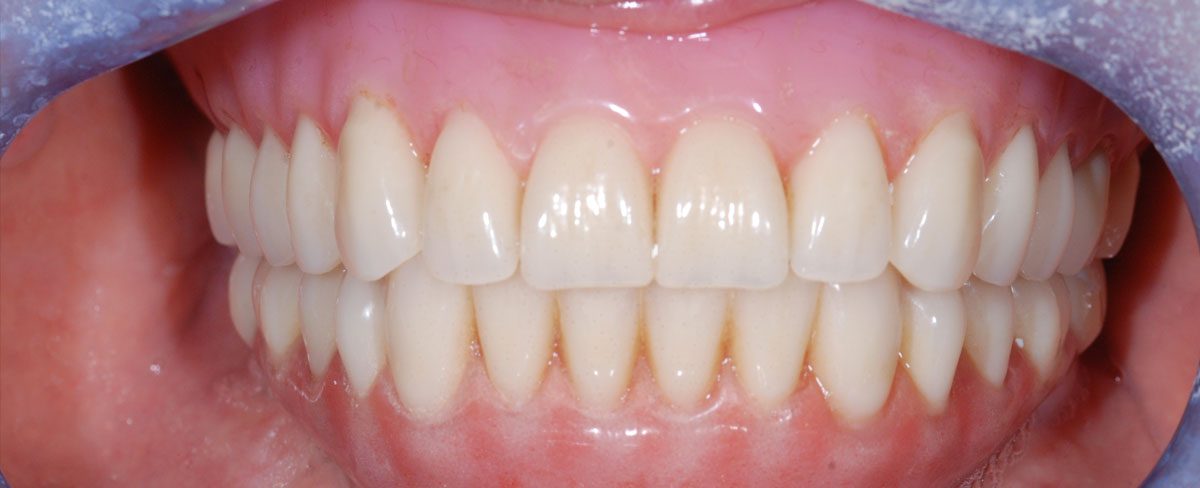
The procedure has a high satisfaction rate, with most patients able to have their dentures fully aligned and attached. A better fit for dental prosthetics results in improved quality of life, especially in dental function, and ensures maximum retention.

How is the Procedure Performed?

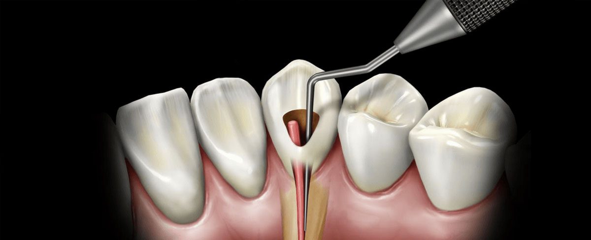
Depending on the need, the patient may be placed under local or general anaesthesia for the procedure. The oral surgeon then makes an incision along the gum line, near the area where trimming is required, and the tissue flap is raised to expose the jawbone. The surgeon then proceeds to remove or drill out unnecessary bone using specialised surgical tools like a rotary drill or chisel.

At this point, there are several techniques that can be employed to remove any extra bone. A simple alveoplasty can be used for the removal of both the buccal alveolar plate and interseptal bone through simple bone trimming.

For patients who have a single tooth extracted, the excess bony tissue on both sides of the socket will be removed. The surgeon will then proceed to remove and file excess bones before ending the procedure.

There are other modifications to these techniques that the surgeon can also perform. These include the radical alveoplasty, which involves the complete removal of the labial plate while another technique requires the removal of the interradicular bone.

After modifying and smoothing out the jawbone, bone particles are removed by washing the surgical area with a saline solution. After making sure that no debris is left behind, the flap is lowered and the incision closed with surgical sutures.


wisdom-tooth-extraction-1200x488.jpg

A wisdom tooth is often extracted to correct an actual problem or to prevent a problem that may come up in the future. When wisdom teeth are starting to erupt, a number of problems can occur: your jaw may not be large enough to accommodate them and they may become impacted and unable to break through your gums. Your wisdom teeth may also break partway through your gums, causing a flap of gum tissue to grow over them. Food and germs can become trapped under the flap and cause your gums to become red, swollen, and painful – these are the signs of an infection. More serious problems can develop from impacted teeth, such as damage to other teeth and bone, or the development of a cyst.

Procedure

An extraction begins with local anaesthetic being placed to numb the tooth and the surrounding area. Once the tooth is completely anaesthetized, the doctor will “wiggle” the tooth in the socket to break the fibrous bonds that hold the tooth to the bone. When the tooth is sufficiently loose, the doctor will use a tool that elevates the tooth out of alignment. Sometimes that will cause the tooth to come out immediately. Often, the doctor will then use the forceps to “grab” the tooth and wiggle it out the rest of the way. Occasionally, if a tooth is more attached to the bone or the roots are misaligned to not allow easy extraction, the doctor will use a drill to cut the tooth into sections. This allows for easier access and less traumatic removal of the tooth.

Extractions are very much about technique and leverage. Our doctors are all taught the latest techniques and have all the equipment needed to look after most extractions. Specific, complicated extractions are often referred to one of the great oral surgeons in the area, if needed. Talk to your dentist about your specific case and the best options for you.


Occlusal-Adjustment-1200x488.jpg

An Occlusal Adjustment procedure or bite adjustment is a procedure performed to remove tiny interferences that keep teeth from coming together properly. The imperfect positioning of the teeth when the jaws are closed is called a malocclusion. These interferences can prevent the jaw from closing in its anatomically correct position. Interferences can occur as teeth develop, after an injury, or accumulate over time with wear.

The following dental treatments can also cause interferences:

  • Fillings
  • Crowns
  • Bridges
  • Extractions

If you suspect that you may need an occlusal adjustment, schedule an appointment with Reedley Family Dental by calling (559) 637-0123.


frenulum-1200x488.jpg

frenectomy (also known as a frenulectomy or frenotomy) is the removal of a frenulum, a small fold of tissue that prevents an organ in the body from moving too far. It can refer to frenula in several places on the human body. It is related to frenuloplasty, a surgical alteration in a frenulum. Done mostly for orthodontic purposes, a frenectomy is either performed inside the middle of upper lip, which is called labial frenectomy, or under the tongue, called lingual frenectomy. Frenectomy is a very common dental procedure that is performed on infants, children, and adults. A similar procedure frenulotomy is where a tight frenulum may be relieved by making an incision in the tight tissue.

There are several frenula that are associated with types of frenectomy:

  • Genital frenectomy can be performed to remove frenulums from genitalia
  • Lingual frenectomy (of the tongue) as treatment for ankyloglossia (tongue-tie)
  • Labial frenectomy (of the lip) is very common with patients undergoing denture treatment in order to get the proper fit of dentures or patients who have tissues attached to centre of upper lip and causing recession of gums or gap between the upper front teeth called central incisors.
  • A frenectomy can also be performed to remove a section of tissue (the frenulum) that attached to the gingival tissue between two teeth.

dental-bone-graft-1200x488.jpg

The main function of a bone graft is that, over a period of time, the jawbone associated with missing teeth atrophies or is reabsorbed. This often leaves a condition in which there is poor quality and quantity of bone suitable for the placement of dental implants or for properly fitting dentures.

A bone graft is a procedure that replaces missing bone with material from the patient’s own body or an artificial, synthetic, or natural substitute. The graft not only replaces missing bone, but it can also preserve, and in some cases, reverse bone loss. Over time, this new growth then strengthens the area by forming a bridge between your existing bone and the grafted material.

Bone grafts and implants give patients the opportunity to replace missing teeth, to gain back their confidence and restore esthetic appearance and functionality.


Copyright 2020 - Reedley Family Dental. Web Design & SEO by Tommy Le Inc.