Arestin-1200x488.jpg

Treat our patients beyond the dental office

ARESTIN® (minocycline HCl) Microspheres, 1mg is indicated as an adjunct to scaling and root planing (SRP) procedures for reduction of pocket depth in patients with adult periodontitis. ARESTIN® may be used as part of a periodontal maintenance program, which includes good oral hygiene and SRP.

Arestin® is a bioresorbable gel containing minocycline (a tetracycline derivative) microspheres. It has been FDA approved for treatment of gum disease after a deep cleaning procedure known as scaling and root planing

At Reedley Family Dental, our dental care doesn’t stop once you leave the office. It continues until you are 100% satisfied. If you have any concerns, please don’t hesitate to call our office in Reedley, CA. Our dental professionals are here and happy to help with any questions you may have.


teeth-scaling-polishing-1200x488.png

You visit the dentist every six months for your dental check-up and professional cleaning, then one visit the dentist tells you that you have gum disease. Gum disease is an inflammation of the gum tissue that could affect the teeth and supporting bone in your mouth. Plaque bacteria, acids and certain foods all contribute to the development of gum disease. Fortunately, two common methods exist to reverse the disease — dental scaling and root planing.

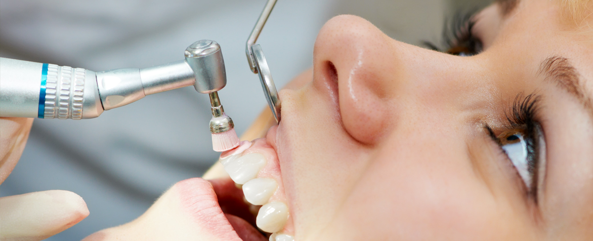
Dental scaling occurs with manual hand instruments, ultrasonic instruments or both. The dentist will start the procedure with a thorough examination of your mouth. Next, an ultrasonic scaling device will be used to eliminate the plaque bacteria with sonic vibrations. The ultrasonic scaling device removes tartar (calculus), plaque and biofilm from the tooth surface and underneath the gum line. A manual instrument may be used next to remove the remainder.

Root planing involves detailed scaling of the root surface to decrease inflammation of the gum tissue. The dentist scales the root surface to smooth out rough target areas, eliminating plaque and biofilm development.

How Will It Feel?

If your gum tissue is sensitive and diseased, local anesthesia can be administered to numb the tissue. If your teeth are sensitive before or after the professional cleaning, it may be recommended that you use a desensitizing paste to provide you with sensitivity relief. Dental scaling and root planing may take two to four visits based upon the level of oral disease in the mouth. If you have periodontal disease, dental scaling and root planing will be completed first and then periodontal surgery will be conducted thereafter.

If you think you may have gum disease, scaling & root planing may just be what you need. Please call Reedley Family Dental at (559) 637-0123 to book a consultation.


dental-sports-mouth-guard-1200x488.png

Grinding your teeth can damage enamel, wear down teeth, cause tooth sensitivity and jaw pain. Studies have shown that grinding can be associated with sleep apnea – a serious medical condition. The noise from teeth grinding and snoring can also disturb your spouse’s sleep if loud enough. Always consult with a dental professional if there are any questions or concerns.

If you clench or grind your teeth you should consider a night guard. This device, which is very similar to a mouth guard worn by athletes, provides a barrier between your top and bottom teeth while you sleep. All night guards are custom fitted for comfort and to allow proper breathing. Your dentist will take an impression of your teeth and have the night guard created by a dental lab. Night guards are very durable and can be used for up to 10 years.

There are also some things that you can do to try to consciously stop teeth grinding. Awareness of the problem can help to identify the times and situations that cause you to grind. Refrain from chewing gum or on other objects like pens. Studies show that you should also avoid drinks with caffeine, as these can increase the likelihood of you grinding your teeth. If you suspect you might be grinding your teeth at night, set up an appointment with us today.

Many people are afflicted with bruxism, or teeth grinding. Some people may do this consciously during the day, but it is a larger problem at night while you are asleep.

Mouth Guards for Sports

A mouth guard should be a top priority in your child’s list of sport equipment. A vacuum-formed custom-fitted mouth guard from our office is your child’s most comfortable and best protection against sports-related injuries.

We’d be happy to prepare a custom sports guard to protect your child.


dental-sealant-1200x488.png

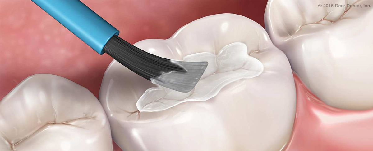
Dental sealant is a thin, plastic coating painted on the chewing surfaces of teeth — usually the back teeth (the premolars and molars) — to prevent tooth decay. The sealant quickly bonds into the depressions and grooves of the teeth, forming a protective shield over the enamel of each tooth.

Although thorough brushing and flossing can remove food particles and plaque from smooth surfaces of teeth, they cannot always get into all the nooks and crannies of the back teeth to remove the food and plaque. Sealants protect these vulnerable areas from tooth decay by “sealing out” plaque and food.

Who Should Get Sealants?

Because of the likelihood of developing decay in the depressions and grooves of the premolars and molars, children and teenagers are candidates for sealants. However, adults without decay or fillings in their molars can also benefit from sealants.

Typically, children should get sealants on their permanent molars and premolars as soon as these teeth come in. In this way, the sealants can protect the teeth through the cavity-prone years of ages 6 to 14.


dentures-1200x488.jpg

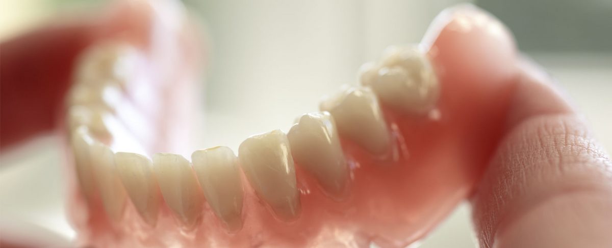
Dentures are a way to replace missing teeth that could be lost due to trauma, decay or advanced gum disease. The main benefit is that you will have a set of fully functioning teeth that are custom fit to you. Today’s dentures are more natural looking and comfortable. Dental technology has advanced with the use of new materials, which means a vastly improved set of dentures. There are two main types of dentures- full and partial. Both types are made in a lab, based on a mold of your mouth.

Traditional Dentures

This is when all the teeth have been removed and the tissue is given time to heal before a full denture is placed. The healing time is usually 6-8 weeks before the gingiva is fully healed. During this time you would be without teeth. This method has not been used as much in recent years.

Immediate Full Dentures

Prior to having your teeth extracted, your dentist will take measurements to have dentures fitted for your mouth. After removing the teeth, the dentures are immediately placed. After the appointment, usually, dentures will be left in your mouth, undisturbed, for 24-48 hours. After that time, you will need a follow-up visit with your dentist to access your healing progress. Adjustments to your denture over the next 4-6 weeks are typical and expected as the mouth heals.

Partial Dentures

This option is available when all teeth do not need to be extracted. A partial denture can replace multiple missing teeth if other options are not feasible.

How to Care for Dentures

Keep your dentures clean! Plaque builds up on dentures just like natural teeth. Unless plaque is removed from your dentures, it can spread to your natural teeth and gums causing gum disease and cavities.

  • Remove your dentures every night.
  • Brush your natural teeth and your gums with a soft toothbrush.
  • Soak your dentures overnight in a special cleaner (a denture cleanser), in warm water, or in a half-and-half mixture of warm water and vinegar. If your dentures have metal clasps, soak in warm water only.

See your dentist regularly because your mouth is always changing, therefore your dentures will need some adjusting from time to time to make sure it fits well. If you have a partial denture, regular check-ups are important to make sure that your natural teeth and gums get the care that is needed.


dental-crowns-1200x488.jpg

Over time, our teeth begin to weaken and become more susceptible to problems such as decay, cracks, discoloration and wear. If you feel that your smile isn’t what it once was, crowns can help you recover your smile. If your dentist notices that a tooth is decayed or seems weakened/cracked, a crown may be necessary to make sure that there are no additional problems with the tooth. In cases like this, a filling or bonding will not be sufficient. Crowns can be made from porcelain, porcelain fused to metal, zirconia, or a full gold crown. A consultation with the dentist is the first step to determine what would be the best option for your particular needs.

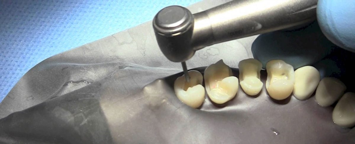
The process of a crown takes two visits to the dentist. On the first visit, the tooth will be reshaped by filing down the enamel so that the crown can be placed over it. You will be given a local anesthetic so that you do not experience any discomfort. Sometimes, if the tooth was broken or the inside of the tooth was compromised, a new filing will be put in to ensure a solid foundation for the crown to sit on. This is called a build up. Once the tooth has been reshaped, an impression will be taken of that tooth and surrounding teeth. This impression will be sent to a dental lab so that your new custom crown can be made. Before leaving the dental office, we will fit you with a temporary crown until your permanent crown is ready.

The crown takes one week to be returned to your dentist. At this time, you will have another appointment to place and fit the permanent crown. You will again have the local anesthetic to numb the area, and the crown will be placed using a cement to ensure that it sets in place. When you look in the mirror, you will see your old smile back. Crowns are durable and will usually last about 10-15 years. You should care for it as you would for your other teeth with regular brushing and flossing.


dental-bridge-1200x488.jpg

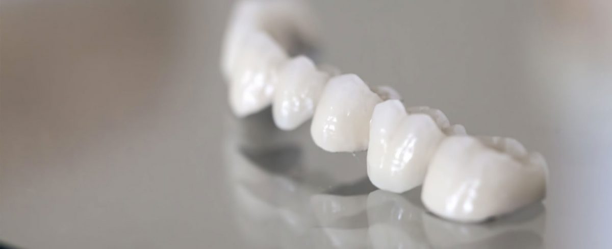
A dental bridge is a restoration that replaces one or more missing teeth. It extends across an area that has no teeth and is typically made up of an artificial tooth fused between two crowns. Bridges are made from gold, metal alloys, porcelain or a combination to ensure that they are strong and durable. The process of creating a bridge begins by creating abutments out of your existing teeth where the bridge will be attached. This process is identical to how crowns are prepared (see crowns). The dentist will often remove and replace any old restorations in these teeth to ensure that there will be no decay present underneath the bridge. These are called build-ups. After the abutments have been created, a mold is taken of the area which is sent to a dental lab. The lab is able to use the mold to create a custom bridge that will fit properly and will feel like your natural teeth. The bridge consists of two crowns on either end to place the abutments, and a pontic – a solid tooth shaped piece – that is the new tooth replacing your missing one.

We will then fit you with a temporary bridge while we wait for the lab to craft your permanent bridge. This will protect the abutment and exposed gum area and look more appealing than having a missing tooth. When the permanent bridge has been created, we will have a follow-up visit to seat the bridge. It will be placed on the abutments, and the dentist will then use an adhesive for the final placement of the bridge.

The bridge may take a while to get used to, but after a few days, it should feel like your own teeth. You should eat soft foods for the first few days. After a short while, you will be able to eat whatever you want with no issues. If you are missing a tooth, you should strongly consider having it replaced, because missing teeth can cause structural changes to your mouth and jaw, as well as making it difficult to eat or speak properly. Set up an appointment today to restore your smile.


Apicoectomy-1200x488.jpg

Your teeth are held in place by roots that extend into your jawbone. Front teeth usually have one root. Other teeth, such as your premolars and molars, have two or more roots. The tip of each root is called the apex. Nerves and blood vessels enter the tooth through the apex, travel through a canal inside the root, and into the pulp chamber, which is inside the crown (the part of the tooth visible in the mouth).

An apicoectomy may be needed when an infection develops or persists after root canal treatment or retreatment. During root canal treatment, the canals are cleaned, and inflamed or infected tissue is removed. Root canals are very complex, with many small branches off the main canal. Sometimes, even after root canal treatment, infected debris can remain in these branches and possibly prevent healing or cause re-infection later. In an apicoectomy, the root tip, or apex, is removed along with the infected tissue. A filling is then placed to seal the end of the root.

An apicoectomy is sometimes called endodontic microsurgery because the procedure is done under an operating microscope.

What It’s Used For

If a root canal becomes infected again after a root canal has been done, it’s often because of a problem near the apex of the root. Your dentist can do an apicoectomy to fix the problem so the tooth doesn’t need to be extracted. An apicoectomy is done only after a tooth has had at least one root canal procedure.

In many cases, a second root canal treatment is considered before an apicoectomy. With advances in technology, dentists often can detect additional canals that were not adequately treated and can clear up the infection by doing a second root canal procedure, thus avoiding the need for an apicoectomy.

An apicoectomy is not the same as a root resection. In a root resection, an entire root is removed, rather than just the tip.

Preparation

Before the procedure, you will have a consultation with your dentist. Your general dentist can do the apicoectomy, but, with the advances in endodontic microsurgery, it is best to be referred to an endodontist.

Your dentist may take X-rays and you may be given an antimicrobial mouth rinse, anti-inflammatory medication and/or antibiotics before the surgery.

If you have high blood pressure or know that you have problems with the epinephrine in local anesthetics, let your dentist know at the consultation. The local anesthetic used for an apicoectomy has about twice as much epinephrine (similar to adrenaline) as the anesthetics used when you get a filling. The extra epinephrine constricts your blood vessels to reduce bleeding near the surgical site so the endodontist can see the root. You may feel your heart rate speed up after you receive the local anesthetic, but this will subside after a few minutes.

How It’s Done

The endodontist will cut and lift the gum away from the tooth so the root is easily accessible. The infected tissue will be removed along with the last few millimeters of the root tip. He or she will use a dye that highlights cracks and fractures in the tooth. If the tooth is cracked or fractured, it may have to be extracted, and the apicoectomy will not continue.

To complete the apicoectomy, 3 to 4 millimeters of the tooth’s canal are cleaned and sealed. The cleaning usually is done under a microscope using ultrasonic instruments. Use of a surgical microscope increases the chances for success because the light and magnification allow the endodontist to see the area better. Your endodontist then will take an X-ray of the area before suturing the tissue back in place.

Most apicoectomies take between 30 to 90 minutes, depending on the location of the tooth and the complexity of the root structure. Procedures on front teeth are generally the shortest. Those on lower molars generally take the longest.

Follow-Up

You will receive instructions from your endodontist about which medications to take and what you can eat or drink. You should ice the area for 10 to 12 hours after the surgery, and rest during that time.

The area may bruise and swell. It may be more swollen the second day after the procedure than the first day. Any pain usually can be controlled with over-the-counter nonsteroidal anti-inflammatory drugs (NSAIDs), such as ibuprofem (Advil, Motrin and others) or prescription medication.

To allow for healing, you should avoid brushing the area, rinsing vigorously, smoking or eating crunchy or hard foods. Do not lift your lip to examine the area, because this can disrupt blood-clot formation and loosen the sutures.

You may have some numbness in the area for days or weeks from the trauma of the surgery. This does not mean that nerves have been damaged. Tell your dentist about any numbness you experience.

Your stitches will be removed 2 to 7 days after the procedure, and all soreness and swelling are usually gone by 14 days after the procedure.

Even though an apicoectomy is considered surgery, many people say that recovering from an apicoectomy is easier than recovering from the original root-canal treatment.

Do you need more information on Apicoectomy (Endodontic Surgery)? Don’t hestitate to call Reedley Family Dental at (559) 637-0123.


root-canal-1200x488.jpg

When the nerve of a tooth is affected by decay or an infection, a root canal procedure is necessary. In order to ensure tooth survival, the living tissue inside the tooth known as the pulp will have to be removed along with the nerves, bacteria, and overall decay. The empty space is then filled with medicated dental materials, specially designed to restore the tooth to full functionality.

Having a root canal performed is the best treatment for saving an infected tooth that would otherwise wither away and die on its own. Many patients believe that removing a tooth is the only way to fix a problem. Not only is this untrue, but removing a tooth can end up being more expensive than other procedures because the infection might spread to adjacent teeth. Root canals are very successful and in most cases, last a lifetime. If there are any new infections that form, the tooth can easily retreat.


Symptoms that might require a Root Canal:

  1. Swelling or tenderness
  2. Heat and cold sensitivity
  3. Severe toothache (worse with pressure)
  4. Presence of an abscess (round bump) on the gums

inlay-onlay-1200x488.jpg

Traditionally, inlays were used instead of fillings to replace a small amount of tooth structure loss due to dental decay. Like fillings, inlays fit inside the tooth.

Dental Inlays

Today, inlays are still being used as dental fillings, but the inlays can be made of a tooth-colored material such as ceramic/porcelain or special dental composite depending on what the patient wants. Defective or unsightly “fillings” can be replaced by tooth-colored inlays and bonded to the tooth in about 30mins to 1 hour. This bonding process may actually improve the strength of the tooth and help seal the inlay to the tooth.

Dental Onlays

Like inlays, onlays also fit inside the tooth, but extend onto the chewing surface of a back tooth to replace one or more cusps. In the past, onlays were made only of gold, but like inlays, more and more patients request a tooth-colored onlay. Making the onlay of ceramic/porcelain allows the restoration to be bonded to the tooth and look natural. This bonding process may actually improve the strength of the tooth and help seal the onlay to the tooth to prevent further decay.


Copyright 2020 - Reedley Family Dental. Web Design & SEO by Tommy Le Inc.